Research
Generating neuronal diversity in fly eyes and human retinal organoids
A central challenge in developmental neurobiology is to understand how the incredible diversity of neuronal cell types is generated. Our lab studies this question in the visual systems of flies and humans. By studying highly divergent organisms, we aim to identify fundamental mechanisms that diversify neuronal function during development.
The random mosaic of photoreceptors in the fly eye
“I, at any rate, am convinced that He does not play dice.”
These famous words were spoken by Albert Einstein about the existence of an underlying reality and the predictability of the universe. A simple look at a pair of twins would back the notion that nature and biology can be amazingly reproducible. However, one would only have to look into the individual cells of these twins to see something remarkable: the different types of cells are sometimes randomly distributed and thus each twin is unique. Only recently have we begun to understand the mechanisms controlling such stochastic developmental events. It appears that noise in molecular processes creates variation, and this variation can be exploited to diversify the functions of cells. Stochastic specification is critical for generating a wide range of cell types, from olfactory neurons that sense odors to B cells required for immune responses. Beyond its role in normal development, stochastic gene regulation can determine whether a person with a mutated gene will suffer from disease. Despite its importance, very little is understood about how this fundamentally different strategy operates.
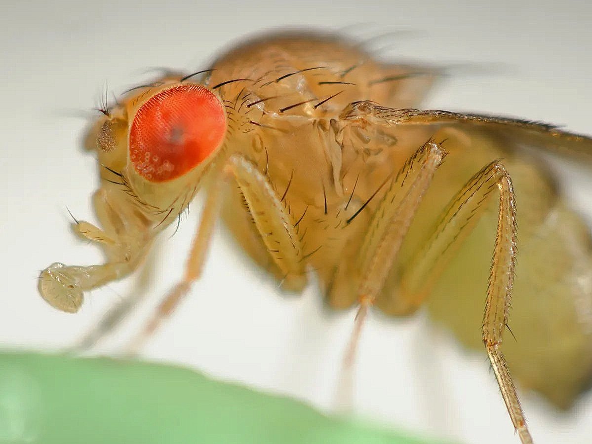
We are using the fly eye as a paradigm to elucidate the mechanisms controlling stochastic gene expression during development. The photoreceptors of the fly eye randomly express light-detecting Rhodopsin proteins. The fly eye is an ideal system to study this phenomenon because it provides a simple binary output for stochastic gene expression, the general mechanisms of cell-fate specification are well-understood, and a vast array of genetic and transgenic tools are available to manipulate cis-regulatory inputs and upstream trans-acting factors.
Project 1. Stochastic photoreceptor specification (Ali Ordway)
The transcription factor Spineless is the critical regulator controlling the random mosaic pattern of photoreceptor subtypes in the fly eye. How does a gene randomly decide to be on or off? To address this question, we are studying how DNA looping, nuclear architecture, chromatin state, and transcriptional regulators control stochastic expression of spineless. We found that a two-step mechanism involving noisy transcription and chromatin compaction determines the on or off state of spineless in mature photoreceptors. This mechanism may represent a general paradigm for gene regulation during development.
Project 2. Nuclear architecture and interchromosomal gene regulation
Very little is known about how long-range interactions of regulatory DNA elements control gene expression. Our studies of nuclear architecture focus on the pairing of homologous chromosomes in somatic cells, which enables gene regulation between chromosomes. We found that chromatin structures called topologically associating domains (TADs) and clusters of DNA-looping insulators button chromosomes together to promote interchromosomal gene regulation during transvection.
The mosaic of photoreceptors in the human eye
The human retina is composed of three cone cell subtypes. These cone cell subtypes are defined by expression of color-detecting opsin proteins: L/red, M/green, and S/blue opsins. Despite their importance in color and daytime vision, very little is understood about how these three photoreceptor subtypes are generated. Since mice and fish have dramatically different patterns of cone cells, we address this question in developing human tissue. We are studying the question of cone cell specification by differentiating human stem cells into retinal organoids. These retinal organoids contain all the cell types of the human retina including opsin-expressing cone photoreceptors (green = L/M opsins; blue = S opsin).
Project: Human cone subtype choice (Christina McNerney and Jo Hagen)
In humans, three subtypes of cone photoreceptors enable trichromatic color and high acuity vision. To overcome the challenges associated with studies of human development, we utilized a human organoid system that recapitulates retinal development and photoreceptor specification. Human stem cell-derived retinal organoids are an ideal system to study cone subtype specification due to their limited number of cone cell subtypes, arrangement in a monolayer for simple quantitative analysis, thin tissue depth allowing live visualization, and tractability for CRISPR-mediated genome engineering. We found that spatiotemporal regulation of thyroid hormone and retinoic acid signaling specifies cone subtypes in human retinal organoids. Our studies advanced human retinal organoids as a model for revealing mechanisms of human development, with promising utility for therapeutics and vision repair.
Retinal ganglion cell (RGC) subtype specification
Retinal ganglion cells are the neurons that connect the retina to the brain. The advent of stem cell-based organoid technology has enabled the interrogation of human biology at the molecular level for the first time. Direct study of developing human tissue will increase our understanding of the mechanisms that control normal organ development, what goes wrong in developmental disorders and disease, and how to manipulate these systems for therapeutic applications. Here, we study retinal organoids to understand the generation and death of RGCs. Elucidating the molecular and developmental mechanisms that specify retinal ganglion cells will be critical for understanding and treating glaucoma.
Project: RGC subtype choice (Brian Guy and Simon Zhang)
We have learned a great deal about the generation of RGCs through the study of model organisms. Human retinal organoids enable, for the first time, the study of RGC specification in developing human tissue. Retinal organoids generate RGCs, yet they are poorly characterized. Our preliminary data suggest that RGCs are the first neurons specified in organoids, consistent with observations in human retinas. Interestingly, recent studies have suggested that there are 20 subtypes of RGCs in humans. Do retinal organoids generate all 20 subtypes? How? If not, why? We are using fluorescence-based cell sorting combined with single cell RNA sequencing to address these questions.


